Rib Cage Muscles Diagram / Intercostal Muscle Strain Physiopedia : It forms the bony framework for breathing.. The human rib cage is made up of 12 paired rib bones; It also protects several vital organs of the chest, such as the heart, aorta, vena cava, and. The sternum, commonly known as the breastbone, is a long, narrow flat bone that serves as the keystone of the rib cage and stabilizes the thoracic skeleton. The function of the rib cage is to filter the blood it receives, processing the blood. As in the typical ribs, the tubercle has a facet for articulation with the transverse process of vertebrae.
Rib cage tendonitis rib cage shape lungs and ribs anatomy rib cage reference female rib cage muscles. Best viewed on 1280 x 768 px resolution in any modern browser. The sternum, commonly known as the breastbone, is a long, narrow flat bone that serves as the keystone of the rib cage and stabilizes the thoracic skeleton. These muscles connect the lower part of the spine to the ilium and the femur and. The rib cage has many attachment points to other important muscles, like the neck, abdominals, and upper back.
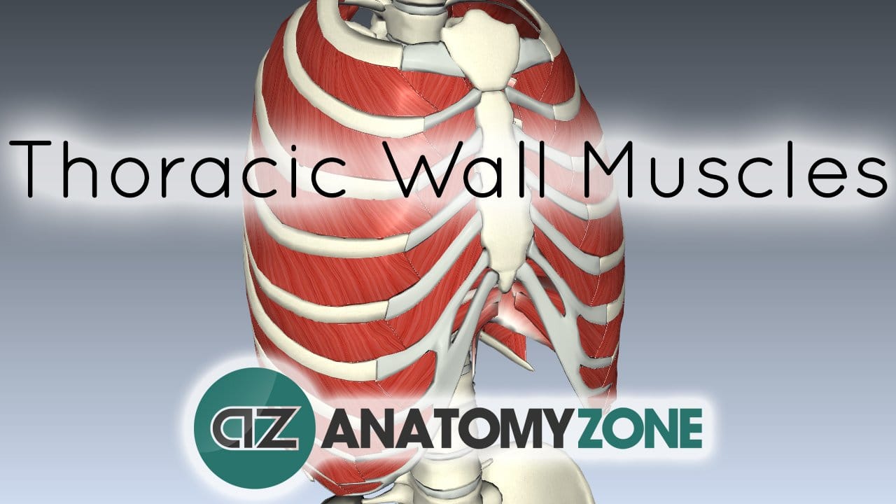
Muscles Of The Thoracic Wall 3d Models Video Tutorials Notes Anatomyzone from anatomyzone.com The rib cage has three important functions: Rib cage pain can be caused by a variety of things, ranging from pulled muscles to a rib fracture. The rib cage muscles consist of the obliques, intercostals and serratus anterior. There are 11 pairs of external intercostal muscles. This is an online quiz called rib cage muscle diagram. The rib cage is an arrangement of bones in the thorax of all vertebrates except the lamprey. In spite of its resistance, the cage is dynamic, allowing pulmonary ventilation to. Intercostal muscle strain is an injury affecting the muscles between two or more ribs.
The rib cage has three important functions:
This muscle helps rotate the upper arm. Anatomy of the rib cage diagram. Start studying rib cage muscles. Related posts of rib cage diagram with organs womens body parts stomach. This is an online quiz called rib cage muscle diagram. As in the typical ribs, the tubercle has a facet for articulation with the transverse process of vertebrae. Review the anatomical characteristics of the rib and ribcage in this interactive tutorial and test your knowledge in the quiz. Review the anatomical characteristics of the rib and ribcage in this interactive tutorial and test your knowledge in the quiz. The dome shaped thoracic cage provides the necessary rigidity for organ protection, weight support for the upper limbs and anchorage for muscles. There are 11 pairs of external intercostal muscles. This is an online quiz called rib cage muscle diagram. It provides a strong framework onto which the muscles of the shoulder girdle, chest, upper abdomen and back can attach. Of all 24 ribs, the first seven pairs are often labeled as 'true.'.
The rib cage is an origin and insertion area for many muscles. The superior surface is unique in that it is marked by two grooves that allow. Intercostal muscle strain is an injury affecting the muscles between two or more ribs. Symptoms of intercostal muscle strain include: The rib cage is made up of the thoracic.
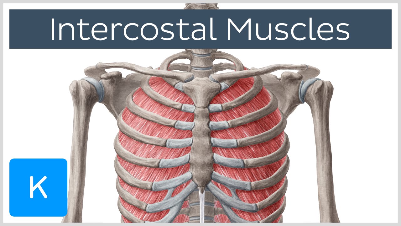
1 Thoracic Wall Radiology Key from i1.wp.com Intercostal muscle strain is an injury affecting the muscles between two or more ribs. The human rib cage is made up of 12 paired rib bones; Review the anatomical characteristics of the rib and ribcage in this interactive tutorial and test your knowledge in the quiz. Several muscles that move the arms, head, and neck have their origins on the sternum. 2006 kia optima belt diagram. In this image, you will find thoracic vertebrum, costochondral joint, costal cartilage, costal margin, costal arch, thoracic vertebrum, xiphoid process, xiphisternal joint, body, manubrial sternal joint, manubrium, the sternal notch in it. It is deep to the erector spinae. They run inferoanteriorly from the rib above to the rib below, and are continuous with the external oblique of the abdomen.
This is an online quiz called rib cage muscle diagram.
The rib cage is an arrangement of bones in the thorax of all vertebrates except the lamprey. Originate at the lower border of the rib, inserting into the superior border of the rib below. The erector spinae group has been ghosted in on the left side. Anatomical body 12 photos of the anatomical body anatomical body diagram, anatomical body picture, anatomical planes and body movement, anatomical position of body, anatomical terms for body parts, human anatomy. This expansion and contraction is facilitated by the intercostal muscles if they are balanced in tone and well aligned posturally. It encloses and protects the heart and lungs. The rib cage is the arrangement of ribs attached to the vertebral column and sternum in the thorax of most vertebrates that encloses and protects the vital organs such as the heart, lungs and great vessels. Of all 24 ribs, the first seven pairs are often labeled as 'true.'. It forms the bony framework for breathing. Rib cage diagram this summary post is displaying rib cage diagram. Human male anatomy, 3/4 figure muscular and skeletal systems, back and front perspective views. The rib cage muscles consist of the obliques, intercostals and serratus anterior. In this image, you will find thoracic vertebrum, costochondral joint, costal cartilage, costal margin, costal arch, thoracic vertebrum, xiphoid process, xiphisternal joint, body, manubrial sternal joint, manubrium, the sternal notch in it.
Muscles that move the rib cage attach to the rib cage. Area between the head and the tubercle of the rib. The pain will get worse when you twist, stretch, breathe in. A group of muscles connected to the rib cage, which help stabilize the shoulder. In this image, you will find thoracic vertebrum, costochondral joint, costal cartilage, costal margin, costal arch, thoracic vertebrum, xiphoid process, xiphisternal joint, body, manubrial sternal joint, manubrium, the sternal notch in it.
Intercostal Muscles Function Area Course Human Anatomy Kenhub Youtube from i.ytimg.com Rib cage pain can be caused by a variety of things, ranging from pulled muscles to a rib fracture. It is deep to the erector spinae. It provides a strong framework onto which the muscles of the shoulder girdle, chest, upper abdomen and back can attach. Human muscles · april 17, 2020. Rib cage diagram this summary post is displaying rib cage diagram. Your intercostal muscles are the muscles between your ribs. The intercostal muscles have different layers that are attached to the ribs to help build the chest wall and. This is an online quiz called rib cage muscle diagram.
The rib cage is composed by sternum, costal cartilages, and ribs connected to the thoracic vertebrae.
Rib cage pain can be caused by a variety of things, ranging from pulled muscles to a rib fracture. Start studying rib cage muscles. The sternum, commonly known as the breastbone, is a long, narrow flat bone that serves as the keystone of the rib cage and stabilizes the thoracic skeleton. Human muscles · april 17, 2020. The intercostal muscles have different layers that are attached to the ribs to help build the chest wall and. The superior surface is unique in that it is marked by two grooves that allow. The function of the rib cage is to filter the blood it receives, processing the blood. Human male anatomy, 3/4 figure muscular and skeletal systems, back and front perspective views. The rib cage has many attachment points to other important muscles, like the neck, abdominals, and upper back. Your intercostal muscles are the muscles between your ribs. There are 11 pairs of external intercostal muscles. Rib cage diagram this summary post is displaying rib cage diagram. The head only articulates with the body of the t1 vertebra and therefore only one articulatory surface is present.
In this image, you will find thoracic vertebrum, costochondral joint, costal cartilage, costal margin, costal arch, thoracic vertebrum, xiphoid process, xiphisternal joint, body, manubrial sternal joint, manubrium, the sternal notch in it rib cage muscles. Our latest youtube film is ready to run.

0 Comments:
Post a Comment